|
Ultrasound (Sonogram)
|
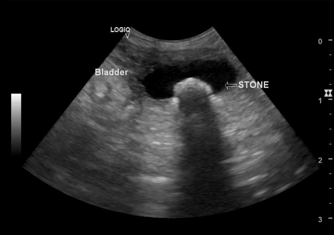
 Ultrasound of bladder with stone (Ultrasound and image by Cinco Animal Hospital). Ultrasound of bladder with stone (Ultrasound and image by Cinco Animal Hospital).
|
Unlike radiographs, no radiation is used in an ultrasound study but instead makes use of sound waves. The ultrasound waves move out from the wand and either become absorbed into organs, pass through them, or are reflected (echo) back. Depending on how many sound waves are absorbed or reflected, an image of the internal organs is formed by a sophisticated integrated computer, and the image is then displayed on a monitor. Real-time moving images are displayed, and still images can be captured as well.
Ultrasound is painless and does not require anesthesia or even sedation in most cases. For an ultrasound evaluation to be done, the pet does need to have the hair shaved from the evaluation area because it will interfere with the images.
|
This test is typically done after blood tests, x-rays, or a physical examination indicating a possible problem. It is useful for evaluating abdominal organs and the reproductive system. As with people, an ultrasound examination can also be used during pregnancies. A specific ultrasound called an echocardiogram is used to visualize the heart and blood vessels as well as its valves.
Ultrasounds are not as good at seeing through air or bone, so it does not replace radiography. It is common to do both x-rays and ultrasound in order to get a good picture of what is going on.
Due to the high cost of the equipment, CT scans and MRIs are more expensive than radiography or ultrasonography, and are usually available only in referral centers or large practices.
Dental Radiographs
|
Dental radiographs are an important diagnostic tool available to veterinary medicine. These images allow to view the internal anatomy of the teeth, the roots, and the bones. Intra-oral radiographs before a scheduled dental cleaning can provide a higher quality of examination of individual teeth or areas of the mouth.
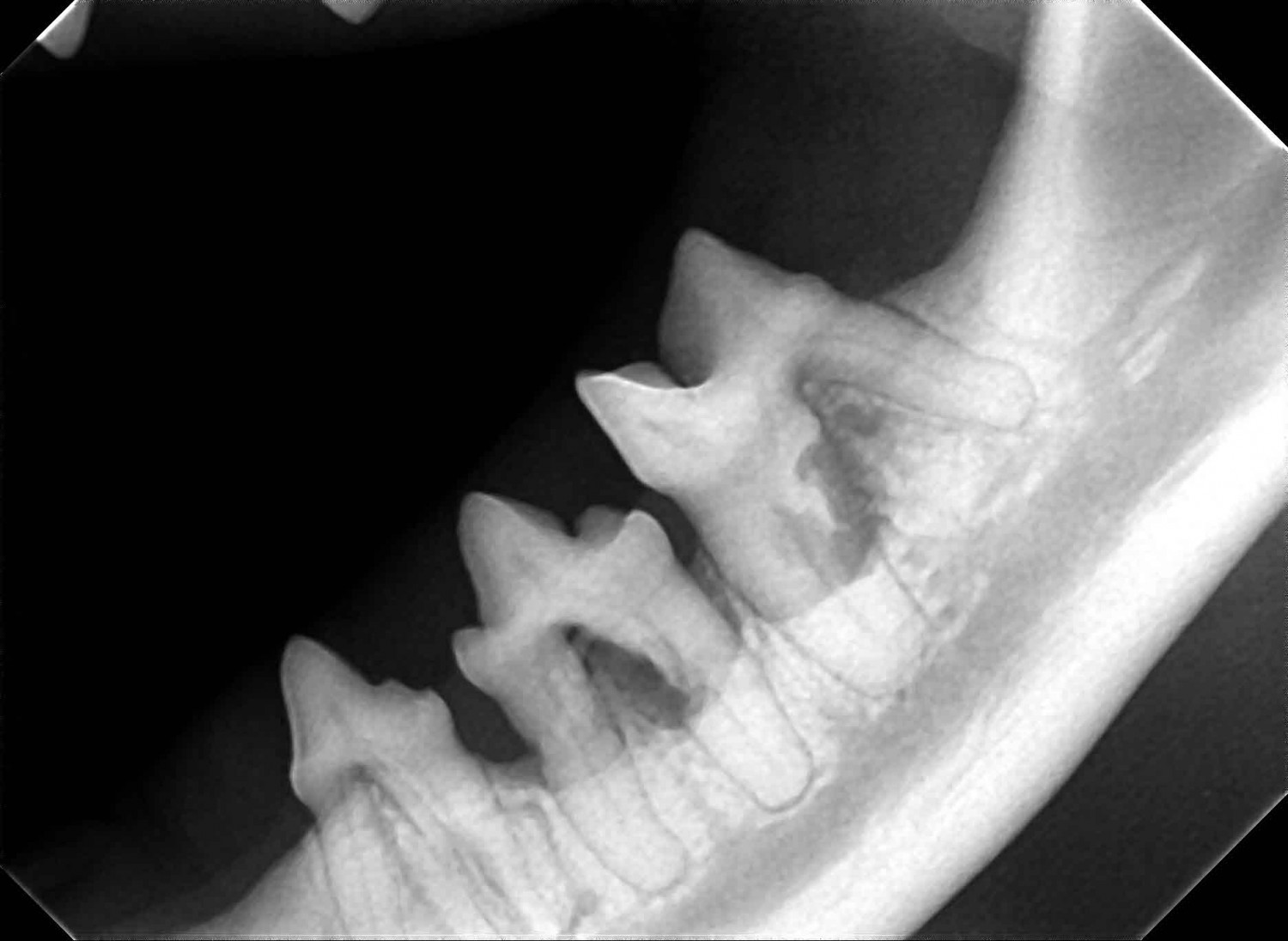
This dental x-ray to the right (Figure 1), there was internal resorption in the mesial root of the molar tooth which was not obvious from the visual exam alone. Without dental x-rays, it is easy to miss these types of painful root lesions, and may have not been taken care of at the time with just a dental cleaning.
|
 Figure 1. Feline internal resorption of the 3rd and 4th mandibular pre-molar and molar tooth Figure 1. Feline internal resorption of the 3rd and 4th mandibular pre-molar and molar tooth
(Image by Cinco Animal Hospital).
|
|
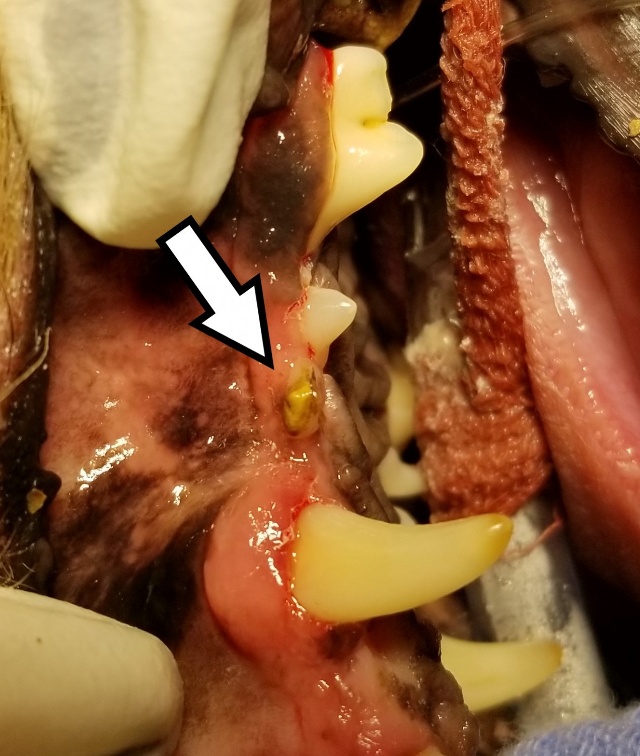
 Figure 2. Without taking radiographs, it appears to be calculus buildup on the tooth Figure 2. Without taking radiographs, it appears to be calculus buildup on the tooth
(Image by Cinco Animal Hospital).
|
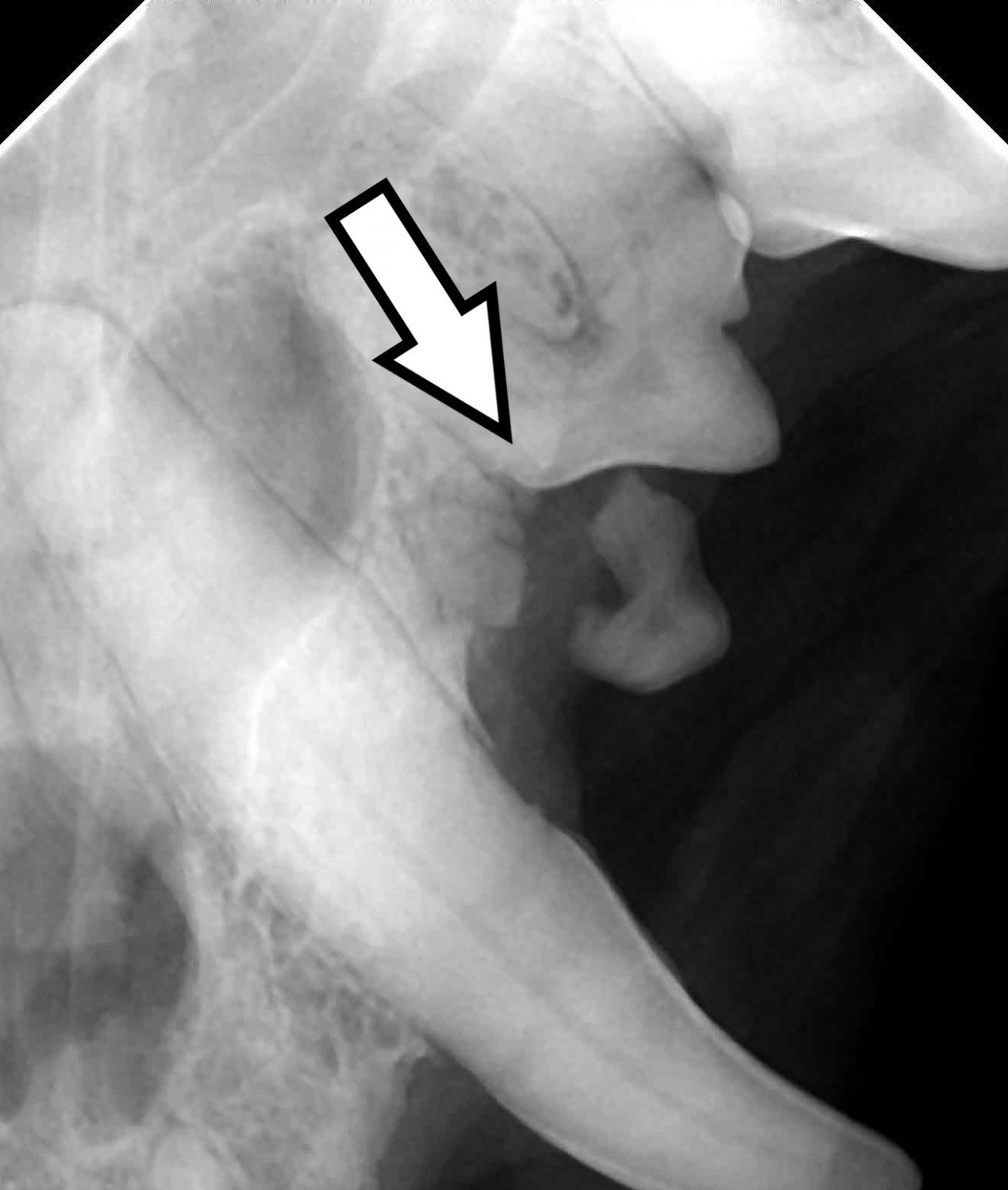
 Figure 3. Radiographs show the same tooth broken under the gum line Figure 3. Radiographs show the same tooth broken under the gum line
(Image by Cinco Animal Hospital).
|
As shown in Figure 2 above, it looks like the tooth has a high build up of dental tartar and gingivitis along the gum-line. Dental radiographs help us visualize the root system for better diagnosis. In Figure 3, we are able to see the tooth has actually broken off from the root and needs to be extracted.
|
